Advances in Intelligent Visual Analysis Methods for Organoids
Editor: | Jun 29,2026
Organoids are important three-dimensional in vitro models for disease modeling, drug screening, precision medicine, and regenerative medicine. They can partially recapitulate tissue structures, functional states, and disease-related phenotypes in vitro. As organoid culture systems and microscopy imaging continue to develop, researchers need stable and reproducible quantitative analysis of organoid growth, morphology, and culture quality. However, bright-field organoid images often contain large morphological variation, blurred boundaries, dense overlap, and complex background interference. These factors make manual observation and measurement inefficient and subjective, and they can also lead to inconsistent judgments among different observers, limiting high-throughput research, drug evaluation, and translational applications.
To address these challenges, Prof. Pengwei Hu’s team at the Xinjiang Technical Institute of Physics and Chemistry, Chinese Academy of Sciences, together with collaborators from Peking University Third Hospital, Chongqing General Hospital of Chongqing University, and Merck KGaA, developed OrgLine, an end-to-end intelligent visual analysis pipeline for organoids. The pipeline is designed for bright-field organoid image analysis and integrates automated detection, instance segmentation, morphometric quantification, time-series tracking, and passaging assistance. Rather than serving as a single-task segmentation tool, OrgLine draws on the general representation ability and prompt-based segmentation concept of large-scale vision models, and adapts them with the team’s pre-trained organoid detection model. It can be viewed as an intelligent visual foundation model for organoid morphometric analysis. It first automatically localizes organoids and generates bounding boxes, then uses these boxes as prompts to guide the segmentation model and obtain precise masks for individual organoids, enabling automated detection, counting, classification, instance segmentation, and morphometric quantification.
This study provides an automated, quantitative, and scalable technical tool for organoid culture and analysis. Through systematic validation using more than 8,000 microscopy images and over 120,000 organoid instances, OrgLine maintained stable performance in scenarios with complex backgrounds, blurred boundaries, and overlapping organoids. Its analysis results were highly consistent with expert manual annotations, helping reduce subjective variation from manual observation. The method can support organoid culture quality control, morphological phenotyping, time-series tracking, and passaging assistance, while enabling non-invasive monitoring of organoid growth based on bright-field microscopy images. It turns experience-based observation into continuous, objective, and comparable data records, supports standardized assessment across different batches and sources of organoids, and provides more reliable quantitative support for drug screening, precision medicine, and high-throughput organoid research.
The research article, entitled “OrgLine: A versatile pipeline for organoid morphometry using detector-guided prompts,” was published in Cell Reports Methods.
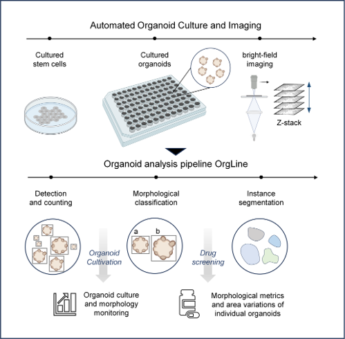
Figure 1. Graphical abstract of OrgLine. The pipeline connects organoid culture and bright-field microscopy with automated detection and counting, morphological classification, instance segmentation, and downstream quantitative analysis.
附件下载:
 86-991-3835823
 lhszhb@ms.xjb.ac.cn
 86-991-3838957
 40-1 Beijing Nan Road, Urumqi, Xinjiang, 830011, China


Copyright @ Xinjiang Techinical Institute of Physics and Chemistry, Chinese Academy of Sciences. All Rights Reserved